Question
A 72-year-old woman is reviewed for a slowly enlarging lesion near the lower eyelid that has been present for many months. It is painless and has gradually developed a shiny raised edge with fine surface vessels and a small area of central ulceration. She has a long history of occupational sun exposure.
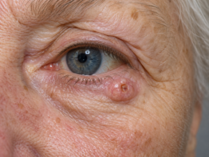
What is the most likely diagnosis?
A. Sebaceous carcinoma
B. Squamous papilloma
C. Chronic blepharitis with granuloma
D. Cutaneous squamous cell carcinoma
E. Basal cell carcinoma
Answer
E. Basal cell carcinoma (BCC)
Explanation
The characteristic description is of a nodular basal cell carcinoma, the commonest form of BCC.
The classic MRCP clues are:
- Sun-exposed area, particularly the head, neck and face
- Slow-growing, usually painless lesion
- Pearly or translucent raised edge
- Telangiectatic vessels over the surface
- Progressive central ulceration, producing the classic “rodent ulcer”
- Mainly local invasion rather than distant spread
- Metastasis is exceptionally rare
BCC arises from basal cells of the epidermis and is the most common skin malignancy. Although it rarely metastasises, untreated lesions can cause considerable local destruction, particularly around anatomically important areas such as the eyelids, nose and ears.
For suspected BCC, appropriate specialist referral is required. Treatment is selected according to the lesion’s site, size, histological subtype and recurrence risk. Surgical excision is commonly used; other selected options include curettage, cryotherapy, topical imiquimod or 5-fluorouracil, and radiotherapy. Lesions in high-risk facial locations may require more specialised surgical approaches such as margin-controlled surgery.
Summary for quick exam revision
Basal cell carcinoma is the commonest skin cancer and usually develops on chronically sun-exposed skin, particularly the head and neck. The classic nodular BCC begins as a slow-growing pearly or flesh-coloured papule. Fine telangiectatic vessels are often visible over its surface. As the lesion enlarges, it may develop central ulceration with a raised or rolled border, producing the classical rodent ulcer appearance. BCC is typically painless and progresses slowly over months or years. Its major clinical behaviour is local invasion and tissue destruction rather than systemic spread. Metastasis is extremely rare. Facial lesions are particularly important because even relatively small tumours can damage structures around the eyes, nose and ears. Long-term ultraviolet exposure is an important risk factor. When BCC is suspected, the patient should be referred for definitive assessment and treatment. Surgical excision is a common definitive treatment. Curettage and cryotherapy may be suitable for selected low-risk lesions. Superficial lesions can sometimes be managed with imiquimod or topical 5-fluorouracil. Radiotherapy is another option in selected patients. For exams, remember the combination sun-exposed site + pearly papule + telangiectasia + rolled edge + central ulceration = basal cell carcinoma.